Chong-bo Zhao,
Xin-yi Li,
Na Wu,
Wei Peng,
Yu-jie Liu,
Chun-jie Wu ![]()
For correspondence:- Chun-jie Wu Email: wucjcdtcm@163.com Tel:+862861801001
Received: 7 July 2015 Accepted: 4 March 2016 Published: 30 April 2016
Citation: Zhao C, Li X, Wu N, Peng W, Liu Y, Wu C. Effect of Arisaema erubescens (Wall) Schott rhizome extract on rheumatoid arthritis. Trop J Pharm Res 2016; 15(4):805-813 doi: 10.4314/tjpr.v15i4.20
© 2016 The authors.
This is an Open Access article that uses a funding model which does not charge readers or their institutions for access and distributed under the terms of the Creative Commons Attribution License (http://creativecommons.org/licenses/by/4.0) and the Budapest Open Access Initiative (http://www.budapestopenaccessinitiative.org/read), which permit unrestricted use, distribution, and reproduction in any medium, provided the original work is properly credited..
Purpose:To investigate the anti-arthritic activity of the water extract of Rhizoma Arisaematis (WERA) using a collagen II -induced arthritis (CIA) rat model.
Methods: CIA was induced in male Sprague-Dawley rats by intradermal injection of bovine collagen II in Complete Freund’s Adjuvant. The rats were treated with daily oral doses of WERA (100, 200, and 400 mg/kg) for 21 consecutive days. Methotrexate (MTX, 3 mg/kg), used as a positive control, was administered orally 2 times/week for 3 weeks. The severity of arthritis was evaluated using indices of paw swelling, arthritic score, body weight, thymus index, and spleen index. In addition, the serum levels of IL-1β, IL-6, IL-10, and TNF-α were measured.
Results: All doses of WERA significantly inhibited paw edema (p < 0.01), decreased arthritis scores (p < 0.01) and spleen index (p < 0.05), and alleviated the weight loss associated with CIA in rats. Furthermore, TNF-α, IL-1β, and IL-6 serum levels were significantly decreased (p < 0.05) by all doses of WERA. By contrast, IL-10 serum levels were markedly increased (p < 0.05).
Conclusion: WERA exerts therapeutic effects in CIA in rats by decreasing the serum levels of TNF-α, IL-1β, IL-6 and IL-10, suggesting WERA may be an effective candidate drug for treating human rheumatoid arthritis.
Introduction
Rheumatoid arthritis (RA) is an autoimmune disease characterized by synovial inflammation and hyperplasia, cartilage and bone destruction, and chronic joint destruction with bone erosion in the extremities (especially the fingers) [1]. In addition, RA may also affect multiple organs and tissues, including the heart, lung, and nervous system. The average morbidity of RA is 1 % throughout the world, and the prevalence of RA is three times higher in females than in males [2]. This disease can rapidly progress into multi-system inflammation with irreversible joint damage, causing premature mortality, disability and compromised quality of life in the industrialized and developing world [3,4]. Currently, the major categories of RA medications used in the clinical setting include disease-modifying anti-rheumatic drugs (DEMARDs), non-steroidal anti-inflammatory drugs (NSAIDs), steroid hormone and biologics (TNF-α antibody and the decoy TNF-α receptor) [5]. Long-term use of these drugs leads to immune system weakness, bone marrow suppression, liver and kidney impairment, gastrointestinal discomfort and cartilage degeneration [6]. Thus, additional effective drugs with low toxicity are needed. In recent years, there have been an increasing number of reports that traditional Chinese medicines (TCMs) can serve as source of new, alternative drugs [7,8]. In addition, most RA patients are very likely to use plant-derived agents for treating RA [9,10]. These reports suggest that finding new RA drugs from commonly used TCMs may be a promising strategy.
Rhizoma Arisaematis (RAM), the tuber of Arisaema erubescens (Wall.) Schott, has been used medicinally for more than two thousand years. As a folk remedy in China, RAM is primarily used to treat rheumatism, swelling, inflammation, and convulsions. In addition, RAM is commonly used as a key component in formulas for treating RA in Chinese folk medicines such as Lijie Capsule, Fufang Nanxing Zhitong Gao, and Aizheng Zhentong Powder [11-14]. Previously published investigations indicated that the active components in RA include alkaloids, flavones, guanosines, polysaccharides, γ-aminobutyric acids, dipeptides, and steroids [15-18].
The aim of the present study was to investigate the anti-arthritic effect of the water extracts of RAM (WERA) and explore its potential mechanisms of action in rats.
Methods
Reagents and apparatus
Bovine collagen type II (CII) and Complete Freund's Adjuvant (CFA) were purchased from Sigma Chemical Co. (Shanghai, China); Methotrexate (MTX) was purchased from Shanghai Sine Pharmaceutical Co., Ltd. (Shanghai, China). TNF-α, IL-1β, IL-6, and IL-10 ELISA kits were purchased from the R&D system (Shanghai, China). All other chemicals and biochemicals used were of the highest grade available.
The following instruments were used: Agilent 1260 Series High Performance Liquid Chromatography-Diode Array Detector (HPLC-DAD); Agilent Technologies, Inc., Folsom, (CA, USA); Plethysmometer PV-200 (ChengDu Technol & Market Co., Chengdu, China); FLx 800 Fluorescence Microplate Reader (BioTek, Shanghai, China).
Preparation of WERA
RAM was purchased from Jiangyou Herbal Medicinal Materials Market (Sichuan Province, China) and identified by Prof. C.J. Wu, Chengdu University of Traditional Chinese Medicine (Chengdu, China). A voucher specimen (S20141015-01) was deposited in the College of Pharmacy of Chengdu University of Traditional Chinese Medicine.
Sliced dried RAM tubers were extracted twice with hot water (each extraction was 2 h). The extracts were centrifuged at 5000 rpm for 20 min to remove starch. The supernatants were dried via a rotary evaporation method at 50 °C to obtain WERA.
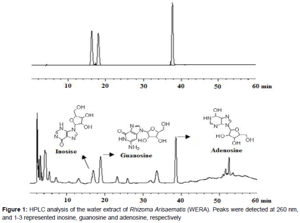
High-performance liquid chromatography (HPLC) analysis of WERA
HPLC separation was performed on the Agilent 1260 HPLC system with a C18 chromatographic column (250 × 4.6 mm, i.d. 5 μm), which was equipped with a quaternary pump, and an automatic thermostatic column compartment. Separation was performed using a gradient elution [H2O (A)/methanol (B)] at a flow rate of 0.8 mL/min. Samples were analyzed using the following gradient program: the run was commenced with 5 % B and then a linear gradient to 10 % B within 6 min, followed by a linear gradient to 20 % B in 30 min that was maintained until 50 min, and finally a linear gradient to 30 % B in 60 min. The sample injection volume was 10 μL, the detection wavelength was set at 260 nm, and the column temperature was set at 30 ºC.
Animals and grouping
Male Sprague-Dawley rats (weighing 160 - 180 g) and KM mice (weighing 18 - 22 g) were purchased from Chengdu Da-Shuo Lab Animal, LTD (Chengdu, China). Animals were kept in an environment with controlled temperature (24 - 26 °C) and photoperiod (12:12 h light–dark cycle) and were given standard commercial rat chow and water. Following 1 week of acclimatization, 10 animals were randomly assigned to each of six groups: the normal group (no treatment), the RA group (no exposure to WERA or MTX) , the MTX group (3 mg/kg, twice/week), the WERA low-dose group (100 mg/kg/d), the WERA middle-dose group (200 mg/kg/d) and the WERA high-dose group (400 mg/kg/d). Animal experiments were conducted in accordance with current ethical regulations for animal care and use at Chengdu University of Traditional Chinese Medicine.
Acute toxicity of WERA
An acute toxicity study of WERA was carried out using Organisation for Economic Co-operation and Development (OECD) guidelines [19]. A series of doses of WERA (5, 50, 500, 2000, 5000, and 10000 mg/kg animal body weight) was prepared by suspending WERA with 0.5 % (w/v) sodium carboxylmethyl cellulose (CMC-Na). Subsequently, the toxicity of WERA was evaluated following oral administration of the prepared series of WERA doses to KM mice. Mortality rates and abnormal behaviors of the mice were observed and recorded within 24 h.
Collagen-induced arthritis (CIA)
The CIA rat model was established according to methods described previously [20]. Bovine CII (2 mg/mL in 0.05 M acetic acid) was emulsified with an equal volume of CFA to achieve a final concentration of 1 mg/mL. Rats were injected subcutaneously with the collagen emulsion (0.5 mg/rat) at the tail root, hind-paw, and three places on the back (dorsonuchal, towards the tail about two-thirds of the way down the back, and towards the tail about one-third of the way down the back). After 1 week, a booster subcutaneous injection of the collagen emulsion (0.5 mg/rat) was administered at the same locations. From days 1 to 21 following the first injection, the WERA and MTX groups were treated orally with WERA and MTX, respectively, and the normal and RA groups were given an equal volumes of CMC-Na.
Determination of arthritic score
The severity of arthritis in CIA rats was assessed according to a previously described method [21, 22]. Paws were examined and graded for severity and loci of erythema, swelling and induration using a 5-point scale (0 = no signs of disease, 1 = signs involving the ankle/wrist, 2 = signs involving the ankle plus tarsal of the hind paw and/or wrist plus carpals of the forepaw, 3 = signs extending to the metatarsals or metacarpals, and 4 = severe disease involving the entire hind or fore paw). The maximum arthritic score per rat was set at 16 (4 points × 4 paws).
Determination of thymus and spleen indices
After 21 days of treatment, the rats were anesthetized with pentobarbital sodium (40 mg/kg, intraperitoneal injection, i.p.) and sacrificed. The thymus and spleen were then promptly removed and weighed. The thymus and spleen indices were expressed as the ratio of the thymus or spleen wet weight (mg/g), respectively, divided by the body weight of the animal [23].
Determination of pro-inflammatory cytokines in serum
Rat blood samples were obtained and allowed to clot for 1h at room temperature. Subsequently, serum was recovered and frozen at -20 °C prior to analysis [24]. The contents of TNF-α, IL-1β, IL-6, and IL-10 in serum were determined by commercial ELISA kits according to the manufacturer’s protocol.
Statistical analysis
Data were presented as mean ±standard deviation and analyzed using SPSS 13.0 statistical software (SPSS Inc., Chicago, IL, USA). Differences between groups were analyzed using one-way analysis of variance (ANOVA), and p < 0.05 was considered statistically significant.
Results
HPLC profile of WERA
The three major constituents in the HPLC fingerprint of WERA were identified as inosine (retention time: 17.237 min), guanosine (retention time: 19.108 min) and adenosine (retention time: 37.762 min). The calculated contents of inosine, guanosine, and adenosine were 0.008, 0.051, and 0.042 mg/g, respectively ().
Acute toxicity of WERA
In accordance with current OECD guidelines, WERA-treated animals were observed individually. Neither death nor any abnormal behaviors were observed during the toxicity study test period. The LD50 value could therefore not be calculated, indicating that oral administration of WERA at doses < 10 g/kg is safe.
Effect of WERA on paw swelling, arthritis score, and body weight
No obvious redness or joint swelling was observed in normal rats (Figures 2 and 3A). However, after CII injection, peripheral paw edema was observed in rats within 24 h. Furthermore, after 21 days of treatment by WERA (200 and 400 mg/kg) and MTX (3 mg/kg), paw edema was significantly decreased (p < 0.01), compared with rats in the RA group (Figures 2 and 3C, E, F).
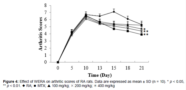
Rat arthritis scores following 21 days of treatment with WERA (200 and 400 mg/kg) and MTX (3 mg/kg) were significantly decreased (p < 0.05, p < 0.01, and p < 0.01, respectively) compared with those of RA rats following 13 days of treatment (). In addition, WERA (100 mg/kg) decreased arthritis scores from the 13th to the 18th day of treatment, but these scores were not significantly different than those for RA rats on the 21st day of treatment.
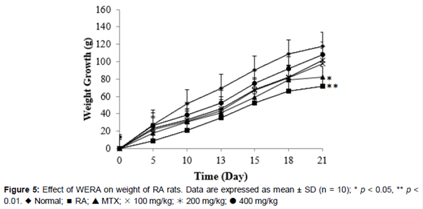
RA rats exhibited marked weight loss compared with normal rats by the 5th day of treatment (p < 0.05; ). During the 2 weeks following the 5th day of treatment, the weight loss of RA rats was reversed by administration of WERA at doses of 100, 200, and 400 mg/kg (p > 0.05). Although WERA-treated rats still showed less weight gain than normal rats, it was higher than the weight gain of MTX-treated rats. Overall, our results demonstrated that animals treated with WERA did not show significant weight loss during the experiment.
Effects of WERA on thymus and spleen indices
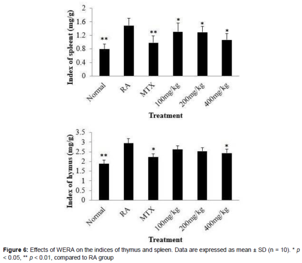
Both thymus and spleen indices in RA group were markedly increased compared with those in the normal group (p < 0.01; ). Interestingly, similar to MTX-treated (3 mg/kg) rats, the spleen indices of WERA-treated rats at doses of 100, 200, and 400 mg/kg were significantly decreased (p < 0.05) compared with those of RA rats. In addition, WERA at 400 mg/kg significantly decrease the thymus index (p < 0.05) compared with that of RA rats.
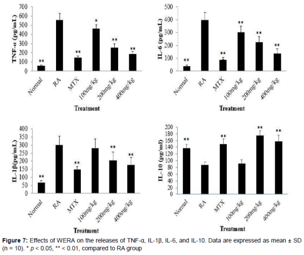
Effects of WERA on serum levels of TNF-α, IL-1β, IL-6, and IL-10
RA rats showed higher serum levels of TNF-α, IL-6, and IL-1β in RA rats (p < 0.01) but lower serum levels of IL-10 (p < 0.05) compared with normal rats (). Similar to MTX treatment (3 mg/kg), WERA treatment (100, 200, and 400 mg/kg) significantly decreased the serum levels of TNF-α (p < 0.05, p < 0.01, and p < 0.01 for the 100, 200, and 400 mg/kg doses, respectively) and IL-6 (p < 0.01) compared with those in RA rats. In addition, WERA (200 and 400 mg/kg) significantly decreased the serum level of IL-1β (p < 0.01) and increased the IL-10 serum level (p < 0.01) compared with those in RA rats.
Discussion
Our investigation confirmed for the first time that WERA can alleviate the joint swelling and inflammatory symptoms of CII-induced RA rats. Our results also indicate that the mechanism of these effects might be a decrease in pro-inflammatory cytokines, including TNF-α, IL-1β, and IL-6.
The CIA animal model reproduces aspects of human RA, from genetic linkage to pathology and clinic manifestations, and is widely used to evaluate the therapeutic effects of candidate drugs and explore the pathogenesis and principles of therapy of RA [26]. Therefore, we used a CIA rat model to evaluate the anti-arthritic activity of WERA. Elimination of clinical symptoms of RA patients is a crucial index for evaluating the therapeutic effects of RA treatment [27], and arthritic scores and paw swelling are two commonly used indices for measurement of the anti-arthritic effects of drugs. Our results show that orally administered WERA significantly relieved joint swelling and redness in RA rats. In addition, treatment with WERA decreased swelling paw volume and arthritis scores and suppressed the weight loss of CII-induced RA rats.
It has been reported that pro-inflammatory cytokines such as TNF-α, IL-1β, and IL-6 play crucial roles in the development of RA [28,29]. TNF-α has been considered to be on top of a cytokine cascade, which can increase the releases of IL-6 and IL-1β, and stimulate cartilage matrix degradation. In addition, IL-1β and IL-6 have been reported to contribute to the development of arthritis [30,31]. IL-10 plays a vitally important role in protecting the integrality of joint tissues and inhibiting releases of pro-inflammatory cytokines in the pathological process of RA [32,33]. Interestingly, in our study, WERA significantly decreased the serum levels of TNF-α, IL-1β, and IL-6, whereas the level of IL-10 was markedly increased. Thus, the potential mechanism of the therapeutic effect of WERA on RA may involve both down-regulating of the level of TNF-α, IL-1β, and IL-6 and up-regulating of the expression of IL-10.
The thymus and spleen are the two major immune organs of body, and their relative weights are usually used to evaluate the immunoregulatory activity of drugs. After administration of WERA, RA rats exhibited a marked decrease of spleen and thymus indices. This result indicated that WERA might affect the immune function on RA rats. HPLC fingerprint analysis showed that the main components of WERA were nucleosides, which were previously found to be immunostimulatory agents [34]. Therefore, our results indicate that nucleosides may be responding active constituents for the anti-arthritic activity of WERA.
Conclusion
The results of this study demonstrate that WERA is effective in treating CII-induced arthritis in rats via decreasing the spleen index, down-regulating TNF-α, IL-1β, and IL-6, and up-regulating L-10. Collectively, the results suggest that Rhizoma Arisaematis can potentially be developed for the treatment or prevention of RA in clinical setting.
References
Archives


News Updates